3D modeling system of blood vessel from medical image
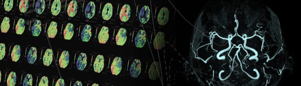
With the rapid development of medical images, it is becoming possible to obtain advanced information on in vivo forms and functions in vivo. Furthermore, by combining a medical image and numerical simulation, it is possible to predict the state after the operation and the progress state of the disease state.
In order to perform numerical simulation, it is necessary to obtain a three-dimensional shape and a center line of a blood vessel from a medical image such as CT (Computed Tomography) or MRI (Magnetic Resonance Imaging), and in general, commercial modeling software is used.
However, this work takes a lot of labor and there is a problem that variations are likely to occur in the shape created by the operator. In addition, when quantitatively comparing three-dimensional shapes obtained from a plurality of medical images, there are many cases that are difficult to solve with commercial modeling software.
Based on the above process, this laboratory is conducting research and development of a three-dimensional shape modeling system for blood vessels (V-Modeler).
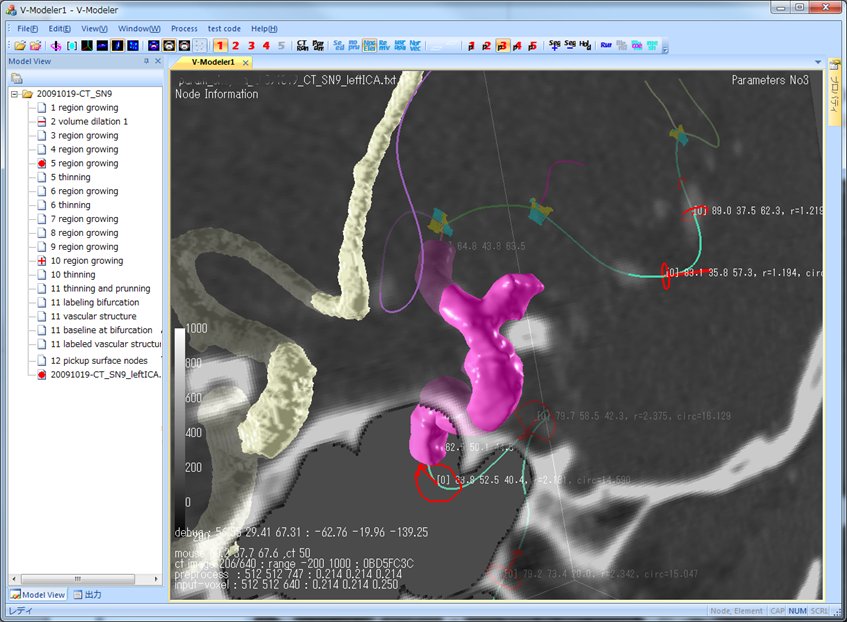
The above figure shows a GUI screen of V-Modeler which is performing three-dimensional shape modeling of a cerebral aneurysm.
Interactive GUI, the efficiency improvement of the three-dimensional shape creation process and the reproducibility of the results, the parameterization of the blood vessel shape using the center line, the formation and measurement of the blood vessel shape for the blood flow simulation, etc. are implemented.
References
[1] 小林匡治,佐藤洋一,大島まり
”医用画像からの血管の3次元形状モデリングの研究開発”
生産研究 64巻3号 pp.319-322(2012)
[2] 小林匡治,佐藤洋一,早川基治,大島まり,
心電同期再構成320列面検出器CT画像を用いた血管壁面トラッキングと血管の材料特性推定に関する研究開発,
第23回バイオエンジニアリング講演会講演論文集,2012,89-90