3D visualization system of 1D-0D blood flow simulation results
In order to predict blood flow changes in the cerebral circulation caused by stent placement for carotid artery stenosis, patient – specific blood flow simulation is a practical technique. Because the blood flow changes with surgical operation, it is necessary to consider the effect of systemic circulation. 1D – 0D blood flow simulation is practical compared with 3D – 1D – 0D blood flow simulation, and it is possible to process patient – specific cerebral circulation taking systemic circulation into account. The purpose of this study is to develop a 1D – 0D blood flow simulation integrated system combined with multimodal medical data such as MRI, CT, SPECT etc which can obtain cerebral circulation information for individual patients.
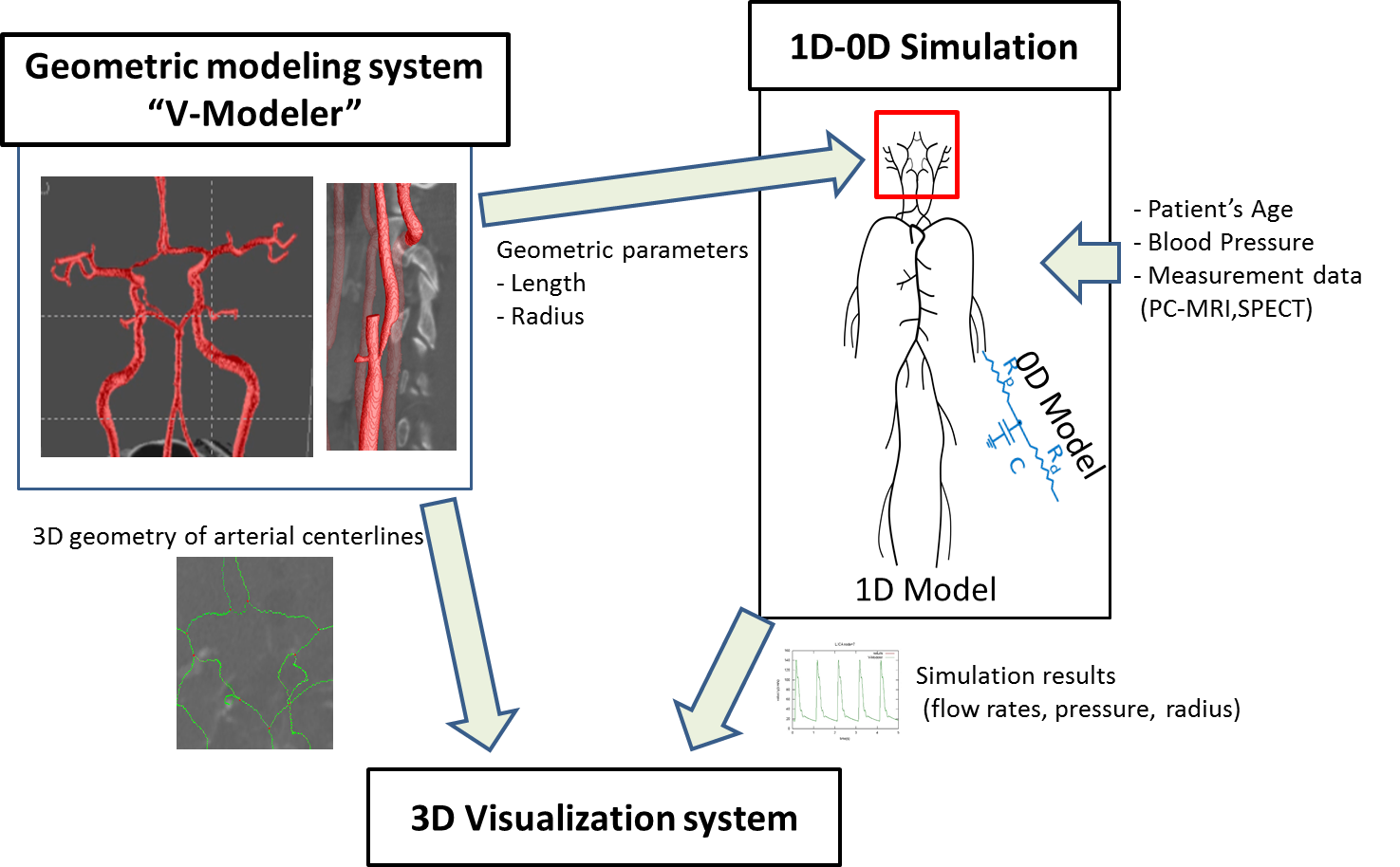
Figure 1.1D – 0D Outline of integrated blood flow simulation system.
As shown in Fig. 1, the integrated system consists of three elements: image based vessel shape modeling system (V – Modeler), 1D – 0D blood flow simulation, 3D visualization system. The blood vessel shape modeling system extracts the blood vessel shape from the medical image as the center line network structure and quantifies the radius and the blood vessel length along the center line. Also in the process of modeling, the stenosis rate is quantified. These quantified data are used in the 1D – 0D blood flow simulation. 1D – 0D blood flow simulation is executed by taking in the patient’s age, blood pressure data, blood flow rate data (PC – MRI, SPECT). The 3D visualization system realizes 3D visualization by mapping the results (flow rate, pressure, radius) of the 1D – 0D blood flow simulation to the center line data obtained from the modeling system
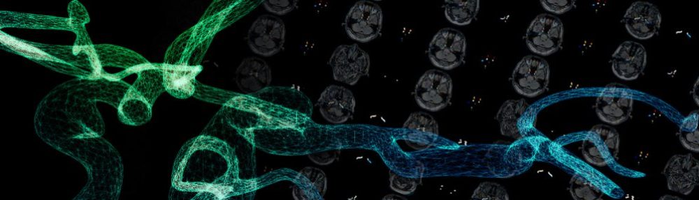
Figure 2. 3D visualization result of 1D-0D blood flow simulation after preoperative operation of stent placement for carotid artery stenosis (blood flow)
Figure 2 shows 3D visualization of the results (blood flow) of 1D-0D blood flow simulation for preoperative and postoperative stent placement for carotid artery stenosis. The blood flow rate of the internal carotid artery before the operation is small in the stenosis side, but it turns out that the left and right are equal after the operation. 1D – 0D We are also studying the verification of the results of blood flow simulation [2].
References
[1] Kobayashi M, Hoshina K, Yamamoto S, Nemoto Y, Akai T, Shigematsu K, Watanabe T, and Oshima M. Development of an Image-Based Modeling System to Investigate Evolutional Geometric Changes of a Stent Graft in an Abdominal Aortic Aneurysm. Circulation advpub, 2015.
[2] Zhang H, Fujiwara N, Kobayashi M, Yamada S, Liang F, Takagi S, Oshima M. Development of a Numerical Method for Patient-Specific Cerebral Circulation Using 1D-0D Simulation of the Entire Cardiovascular System with SPECT Data. Ann Biomed Eng. pp 1-13. 2015 Dec 31.
[3] Kobayashi M, Harada A, Zhang H, Yamada S, Oishi M, Oshima M. Development of an integrated 1D-0D simulation system for patient specific cerebral circulation. Interdiciplinary Cerebrovascular Symposium Intracranial Stent Meeting (ICS2015), 2015.11.13-14.